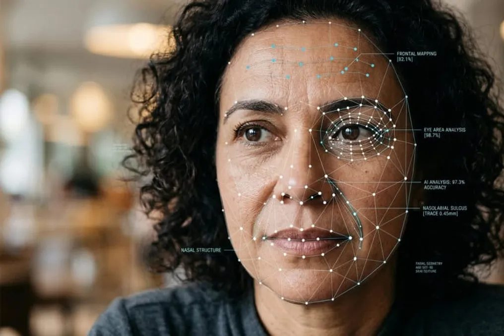
Researchers from the Mass General Brigham healthcare system in the United States have developed an artificial intelligence capable of assessing biological age and predicting the survival probability of cancer patients using only a facial photograph.
The study results, published in The Lancet Digital Health, demonstrate how this analysis can assist doctors in evaluating patient condition before prescribing intensive treatments. However, the tool still requires further research for widespread clinical application.
The system was trained on over 40 million images of faces from people of various ethnic groups and ages. Two separate tools were created from this model. One, FAHR-FaceAge, assesses the biological age of the person depicted and uses this information to determine the prognosis for oncology patients. The second tool, FAHR-FaceSurvival, directly predicts survival chances from the image without relying on age assessment.
Each of these tools focuses on specific areas of the face. FAHR-FaceAge analyzes wrinkles between the nose and the corners of the mouth, as well as the forehead and temple area. Meanwhile, FAHR-FaceSurvival pays attention to the under-eye area and the area above the bridge of the nose.
“As far as we know, this is the first demonstration that an AI model can directly predict cancer survival from a simple picture,” stated Hugo Aerts, one of the lead authors of the study.
To test the model, researchers studied cancer patients who underwent radiation therapy in the United States. Over an average four-year follow-up, it was found that patients who appeared five years older than their actual age had a 21% higher risk of death compared to the other participants. Conversely, the group that looked five years younger had a 17% lower risk.
These data indicate a link between the estimated AI age and survival, but they do not prove that appearance itself determines treatment outcomes.
The study also revealed correlations between the age aging assessed by the system and lifestyle, including certain habits.
Another significant finding was that FAHR-FaceAge maintained consistent results despite various changes in the photographs, such as lighting, facial position, expression, and makeup. Performance also remained stable in most cases of plastic surgery, although a slight change was observed after rhinoplasty.
This technology could potentially provide additional information when assessing patients before surgeries, radiation therapy, and other cancer treatments. It may also help in predicting the risk of complications after major procedures.
Raymond Mac, an oncologist specializing in radiation therapy at Mass General Brigham, explained that the goal is to support individualized medical decisions. According to him, specialists need more advanced methods to assess patients' physical condition before prescribing intensive treatment.
Nevertheless, clinical application still depends on further research. While obtaining a photo is simple, the results must be verified before facial analysis can become part of treatment decision-making.